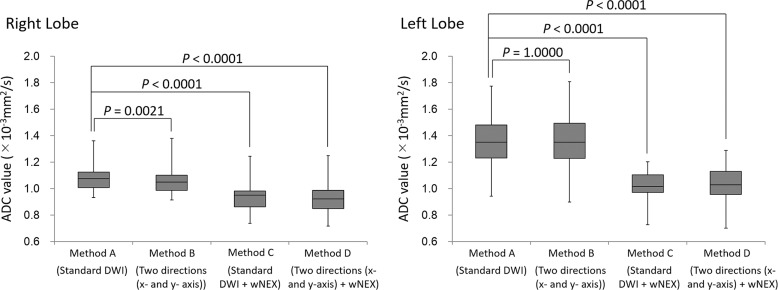
Fig. 6.
Box-plots of the ADC of the right and left lobes in the application study. The ADC for the right lobe of the liver of Method A was significantly higher than those of other methods. The ADC for the left lobe of the liver decreased significantly during acquisition with weighted NEX (Methods C and D). There was no significant difference in the ADC between Methods A and B. ADC, apparent diffusion coefficient; DWI, diffusion-weighted imaging; wNEX, weighted averaging number of excitations.

