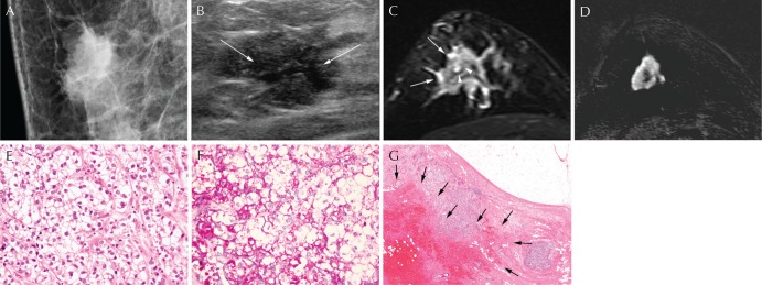
Fig. 1.
A 57-year-old woman with glycogen-rich clear cell carcinoma (GRCC) in the right breast. (A) Mediolateral oblique view of mammography shows an irregular, spiculated, and hyperdense mass in the upper central quadrant of the right breast. (B) Ultrasonography presents an irregular, spiculated, and hypoechoic mass with internal cystic change in the central portion (arrows). (C) On T2-weighted MRI, the mass shows intermediate to low signal intensity with peritumoral edema (arrows) and internal high signal intensity (arrowheads), suggesting cystic portion. (D) The tumor is shown as an irregular, spiculated mass with rim enhancement on subtracted images after contrast injection. (E) The tumor cells shows polygonal contour with clear cytoplasm and distinct cell membrane (Hematoxylin and eosin [H&E] stain ×400). (F) Intracytoplasmic glycogen is highlighted by Periodic acid-Schiff stain (PAS stain, ×400). (G) The tumor mass showed internal cystic portion and hemorrhage (arrows) (H&E stain, ×17).

