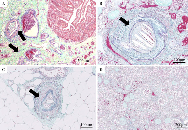
Figure 4.
(A) Surgical specimen of the small intestine resected on day 60. Cholesterol crystal clefts occluded arterioles (arrow). (B, D) Elastica Masson-Goldner (EMG) staining of the kidney from the autopsy specimen. (C) EMG staining of the toe skin from the autopsy specimen. Cholesterol crystal clefts occluding arterioles (arrow).

