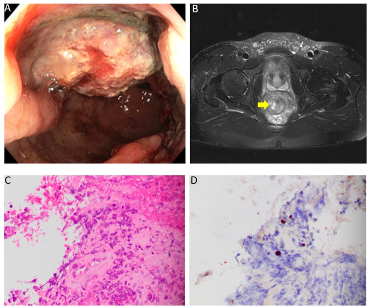
Figure 1.
(A) Colonoscopy showing a fungating and infiltrative partially obstructing medium-sized mass in the rectum, 10–15 cm from the anal verge. (B) Axial T2-weighted MRI showing a polypoidal mass with a circumferential nodular border. More than 50% of the tumor had a very high T2 signal intensity compared to perirectal fat. (C) Histologic sections of the rectal mass biopsy demonstrate colorectal mucosa with ulceration, epithelial cells with nuclear inclusions, exuberant granulation tissue, marked lymphoplasmacytic and eosinophilic infiltrate, and fibrinopurulent exudate (Hematoxylin & Eosin, 400X). (D) Immunohistochemistry for herpes simplex virus-1 was positive in epithelial cells demonstrating a viral cytopathic effect (Herpes simplex virus-1 (HSV-1) immunohistochemistry, 400X).

