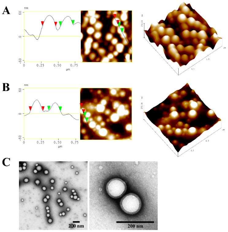
Figure 1.
Morphology of representative curcumin-loaded poly(lactic-co-glycolic acid) (PLGA) nanoparticles (CUR-NP) shown by atomic force microscopy (AFM) (A,B) and transmission electron microscopy (TEM) (C). (A) Freshly prepared nanoparticles, (B,C) nanoparticles post lyophilisation. All AFM micrographs are presented in height mode. Samples were negatively stained with 2% uranyl acetate prior to TEM measurement. Scale bars in TEM micrographs represent 200 nm.

