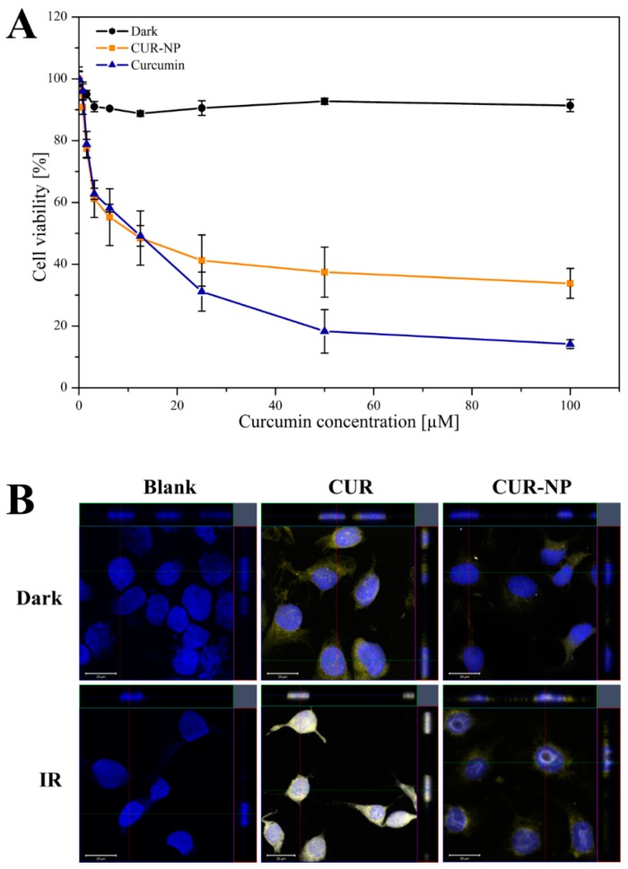
Figure 3.
Phototoxic effect of curcumin-loaded poly(lactic-co-glycolic acid) PLGA nanoparticles (CUR-NP) and free curcumin dissolved in DMSO (CUR) on SK-OV-3 ovarian carcinoma cells: (A) for the MTT-assay, either the nanoformulation or free curcumin was incubated for 4 h at 37 °C and were irradiated at 457 nm with a radiation fluence of 8.6 J/cm2. Dark was used as negative control and represents cells without irradiation. All samples contain 0.1 mg/mL curcumin and were measured in triplicates (n = 9, independent formulations). Results are expressed as means ± SD. (B) CLSM micrographs of SK-OV-3 cells incubated with CUR-NP or free curcumin for 4 h at 37 °C and subsequent irradiation (457 nm, 8.6 J/cm2). The cell nucleus was counterstained with 0.1 µg/mL DAPI and was fixed with 4% formaldehyde solution. The curcumin concentration in CUR-NP was 50 µM. Nuclear damage is clearly witnessed in the irradiated samples whereas in the dark, the nucleus is intact. Dark was used as negative control and represents cells without irradiation. Scale bars denote 20 µm.

