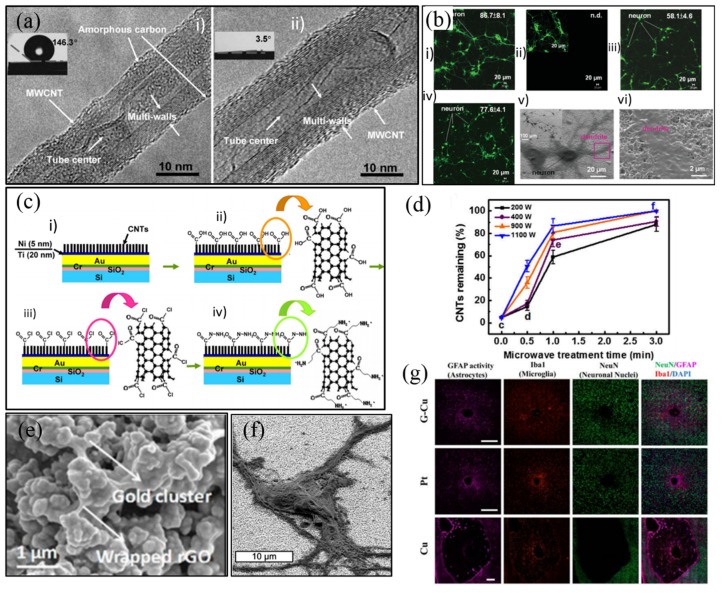
Figure 11.
(a) Transmission electron microscope (TEM) of multi-walled carbon nanotube (MWCNT) with amorphous carbon before and after plasma treatment [115]; (b) fluorescent images of neuron cells cultured on as-grown carbon nanotubes (CNTs) and UV-ozone-modified CNTs, and corresponding SEM images [117]; (c) Process flow of MWCNT amino-functionalization [118]; (d) the percentages of CNTs remaining after 5 min sonication vs. microwave treatment time at various powers [123]; (e) SEM image of reduced graphene oxide (GO) coating [124]; (f) SEM images of neural cells attached well to GO coating [125]; (g) histological studies of tissue response to GO coating [122]. Reproduced from the mentioned references with permission from the related journals.

