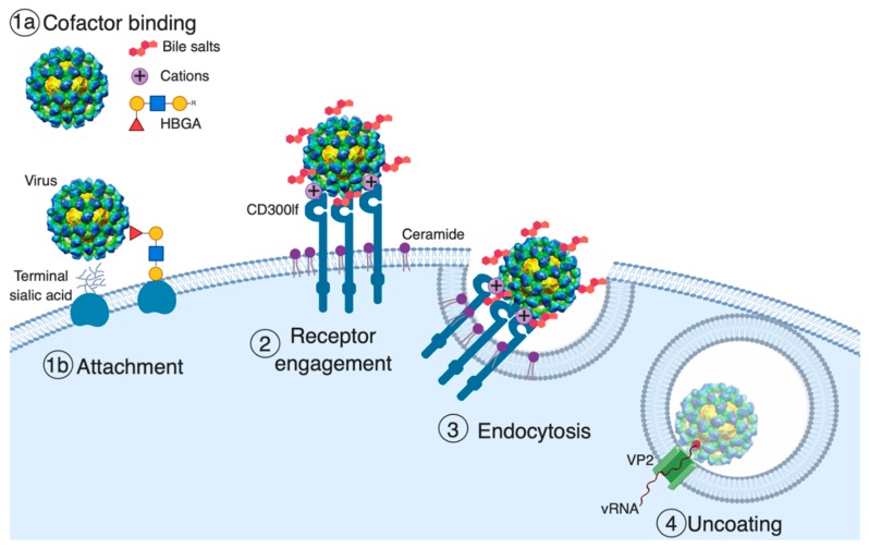
Figure 1.
Model of norovirus entry. The first and often rate-limiting step of viral entry is viral attachment to the cell surface. Cell-associated host glycans including terminal sialic acid and histo-blood group antigens (HBGAs) can facilitate the entry of mouse (MNoV) and human norovirus (HNoV), respectively [37,39,40,41]. Soluble cofactors including soluble forms of HBGAs (HNoV), bile salts (MNoV and HNoV), and divalent cations (MNoV) can also augment the attachment of the virus to cells [25,26,31,38,41]. For MNoV, these soluble cofactors increase virus attachment in a receptor-dependent manner. The second stage of viral entry is receptor engagement. CD300lf, an immunoglobulin (Ig) domain-containing membrane protein, is the MNoV receptor, and feline junctional adhesion molecule A (fJAM-A) is the feline calicivirus (FCV) receptor, while the HNoV receptor remains unknown [26,33,42]. Interestingly, ceramide alters CD300lf conformation or clustering, promoting the interaction with MNoV. Following receptor engagement, the virus is endocytosed where, at least for FCV, receptor binding triggers the minor capsid protein VP2 to form a membrane portal that may enable viral genome release in the cytosol [34].

