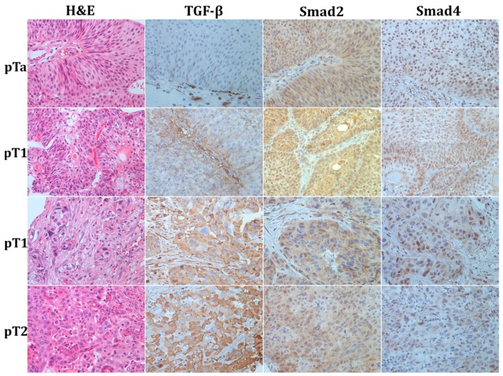
Figure 1.
Representative photomicrographs of haematoxylin–eosin stain and immunohistochemical staining to TGF-β1, Smad2, and Smad4 in urothelial bladder cancer; first row—papillary non-invasive low grade tumor (pTa); second row—superficially invasive low grade tumor (pT1); third row—superficially invasive high-grade tumor (pT1); fourth row—muscle-invasive urothelial bladder cancer (pT2). Original magnification ×400.

