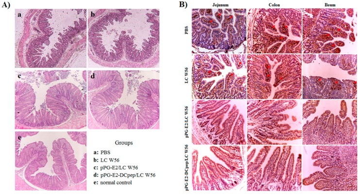
Figure 9.
Histopathological changes (A) and immunohistochemistry (B) observed in the intestine of the mice from pPG-E2-DCpep/LC W56 group, pPG-E2/LC W56 group, LC W56 group, and PBS group on days 15 post-challenge with BVDV. Results showed that no abnormal histopathological changes were observed in mice orally immunized with the pPG-E2-DCpep/LC W56 and pPG-E2/LC W56, whereas prominent histopathological changes, including severe disruption of the intestinal structural integrity and shortening of the villi, developed in mice of LC W56 group and PBS group. Moreover, the immunohistochemical results of the intestine showed that large amounts of virus was observed in the jejunum, colon, and ileum of the mice in PBS group and LC W56 group on day 15 post-challenge, while no virus was detected in pPG-E2-DCpep/LC W56 group and pPG-E2/LC W56 group.

