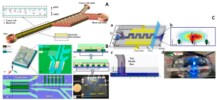
Figure 3.
2D electrode microfluidic chips with DEP. (A) Schematic of the device to separate cancer cells from blood cells by FDEP generated by parallel electrodes. Reproduced with permission from [52], © 2016 John Wiley and Sons. (B) The device combined with induced-charge electro-osmosis (ICEO) for particle focusing and DEP achieving simpler chip structure to perform particle focusing and separation. Reproduced with permission from [60], © 2017 American Chemical Society. (C) Hybrid DEP-inertial device with top sheath flow to improve the separation efficiency. Reproduced with permission from [64], © 2018 Elsevier.

