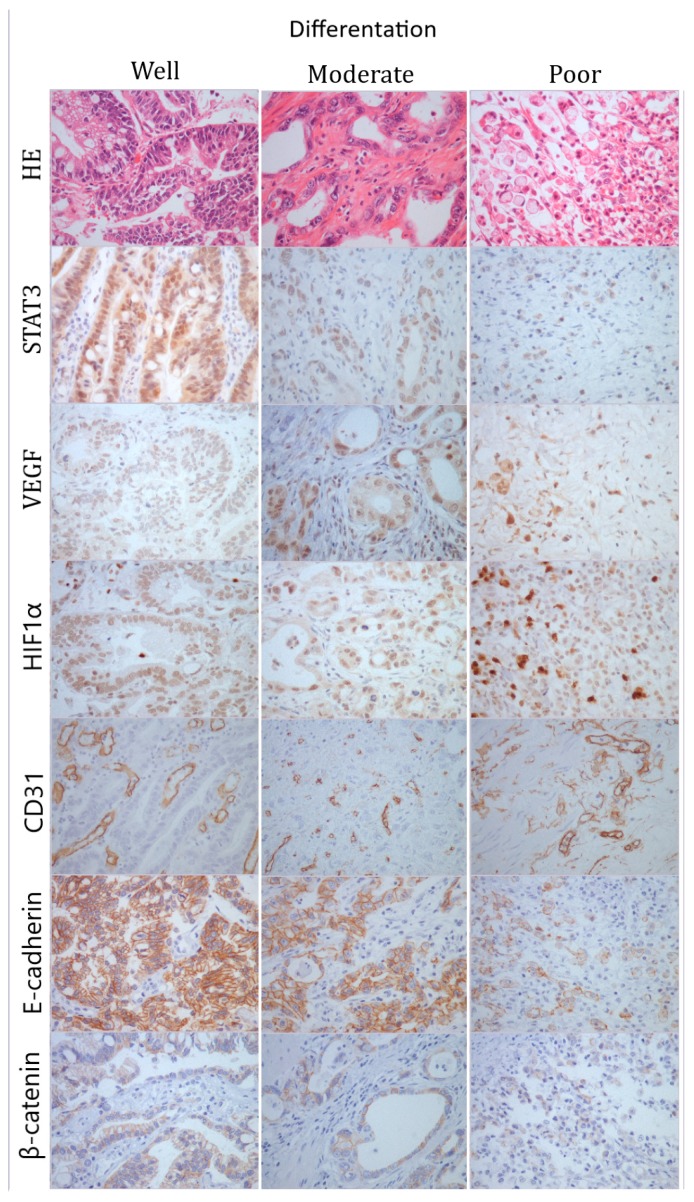
Figure 1.
Representative photomicrographs of HE and immunohistochemical staining to STAT3, VEGF, HIF1α, CD31, E-cadherin, and β-catenin in gastric cancer; first column–well-differentiated tumor; second column–moderately differentiated tumor; third column–poorly differentiated tumor. Original magnification ×400.

