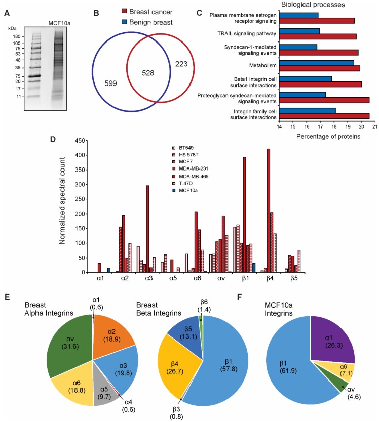
Figure 3.
Breast cancer EV integrin profiles differ from benign breast cell-derived vesicles. (A) Coomassie-stained gel purification of MCF10a EV proteins; (B) Overlap of total vesicle proteins identified by mass spectrometry from MCF10a cells (benign breast) compared to those commonly identified in all six breast cancer cell lines in the NCI-60 panel; (C) Enrichment analysis of proteins identified in benign breast EVs versus breast cancer cell-derived EVs; (D) Spectral count comparison of the most abundant integrin subunits secreted by benign (blue) or tumor (red) breast cells; (E) Breakdown of EV alpha and beta integrin subunit composition (percentage of total alpha or beta proteins, respectively) secreted from breast cancer cells; (F) Vesicle composition of integrin subunits secreted by MCF10a breast epithelial cells (percentage of total integrins identified in samples).

