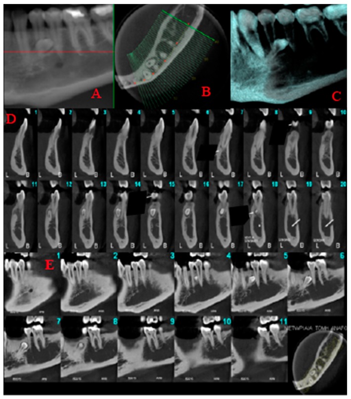
Figure 5.
Limited field of view cone beam computed tomography images. Reformatted panoramic (A), axial (B), volumetric rendering (C) and sequential 1 mm thick/1 mm interval cross-sectional (D) and sagittal (E) cone beam computed tomography images of the left mandible demonstrating the supernumerary tooth, which is lingually located in the root of the lower left first premolar and is tangent to the latter.

