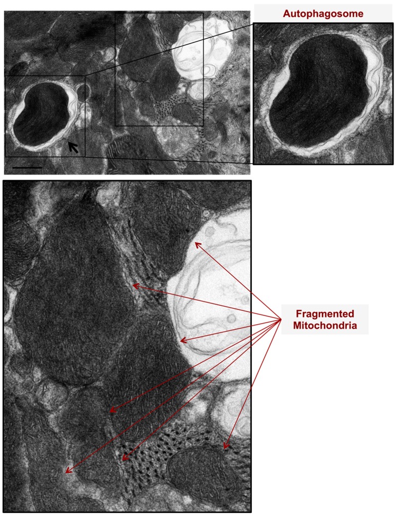
Figure 1.
Mitochondrial autophagy (mitophagy) and mitochondrial fragmentation are pathological features evident in phenylephrine-stressed cardiac myocytes, in vitro. Cardiac myocytes were stressed with 10 μM Phenylephrine for 2 h. Note the presence of autophagosomes, double membranes, and surrounding damaged and fragmented mitochondria (black arrows). Also, note the presence of fragmented mitochondria (red arrows). Photomicrograph is 40k × magnified, scale bar 0.5 μm.

