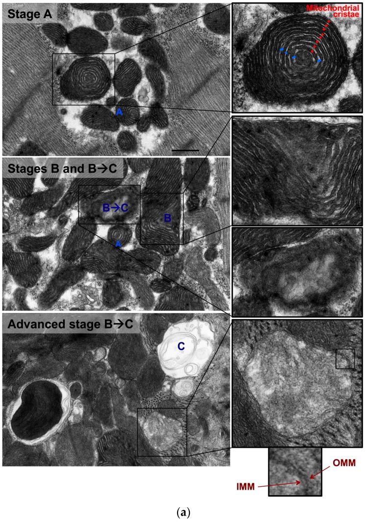
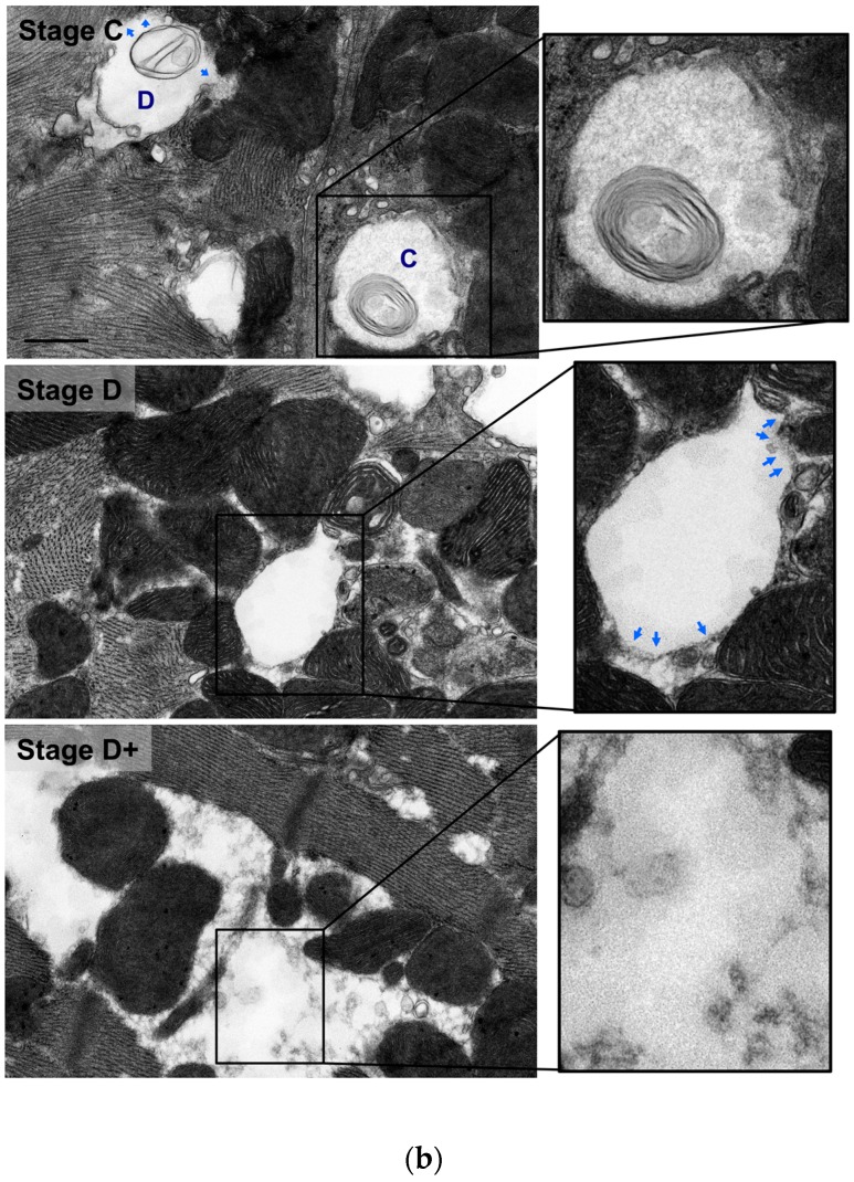
Figure 2.
Morphological stages of mitochondrial vacuolar degeneration in phenylephrine-stressed cardiac myocytes in vitro. Stage A: Highlights concentric, onion-like arrangement of mitochondrion cristae (small red dots) with the widening of spaces between adjacent cristae (blue bars). Stage B: Dissolution of mitochondrion cristae in one mitochondrion area. Stage B→C: (a) Early dissolution of mitochondrion cristae in multiple mitochondrion areas. (b) Advanced stage B→C, mitochondrial swelling with almost complete dissolution of mitochondrial cristae. The inner mitochondrial membrane (IMM) and outer mitochondrial membrane (OMM) remain intact. Stage C: Mitochondrial swelling with the entire dissolution of mitochondrion cristae and the IMM. The OMM remains intact. Stage D: Stage C with an associated rupture of OMM (blue arrows). Stage D+: Area(s) where numerous adjacent mitochondria had undergone complete vacuolar degeneration and the entire dissolution of mitochondrial cristae, IMM and OMM. Photomicrographs are 40k × magnified, scale bar 0.5 μm.


