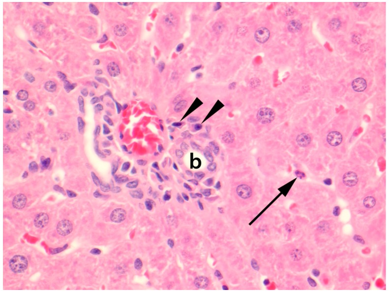
Figure 3.
Photomicrograph of liver parenchyma of a natural Seoul virus (SEOV) infection in a feeder rat. A polymorphonuclear leukocyte (arrow) is present within a hepatic sinusoid, and centrally, a portal area with a bile duct (b) and blood vessels contains a mild lymphoplasmacytic aggregate of which plasma cells (arrowheads) are evident. Stained with haematoxylin & eosin (HE). Original magnification 400×.

