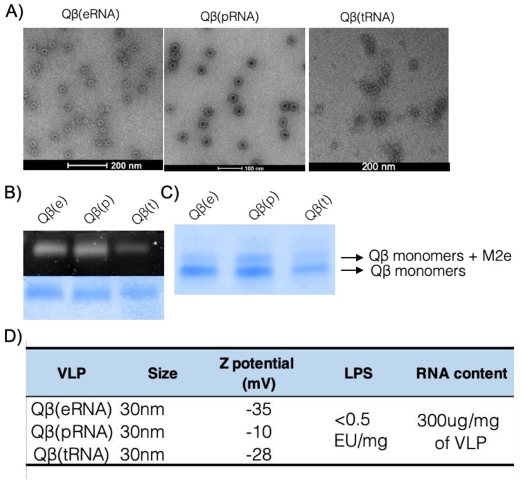
Figure 1.
Vaccine preparation and characterisation. (A) Electron micrograph of re-assembled VLPs. Qβ VLP containing eukaryotic RNA (eRNA), prokaryotic RNA (pRNA) and tRNA (tRNA) have similar structure and size. (B) Native analysis of RNA and protein content of re-assembled VLPs. Upper panel showing SYBR staining and bottom panel showing protein staining of same agarose gel. (C) Similar coupling efficiency of different VLPs and M2 peptides on a reduced SDS-PAGE gel electrophoresis. (D) Characterisation of VLPs. Size was taken as an average of values obtained in the EM images. Z potential was measured by DLS and is expressed as mV. LPS content expressed on endotoxin units (EU) per milligram of VLP. Average RNA content per mg of VLP. Results are representative of more than 5 independent purification and re-assembly studies.

