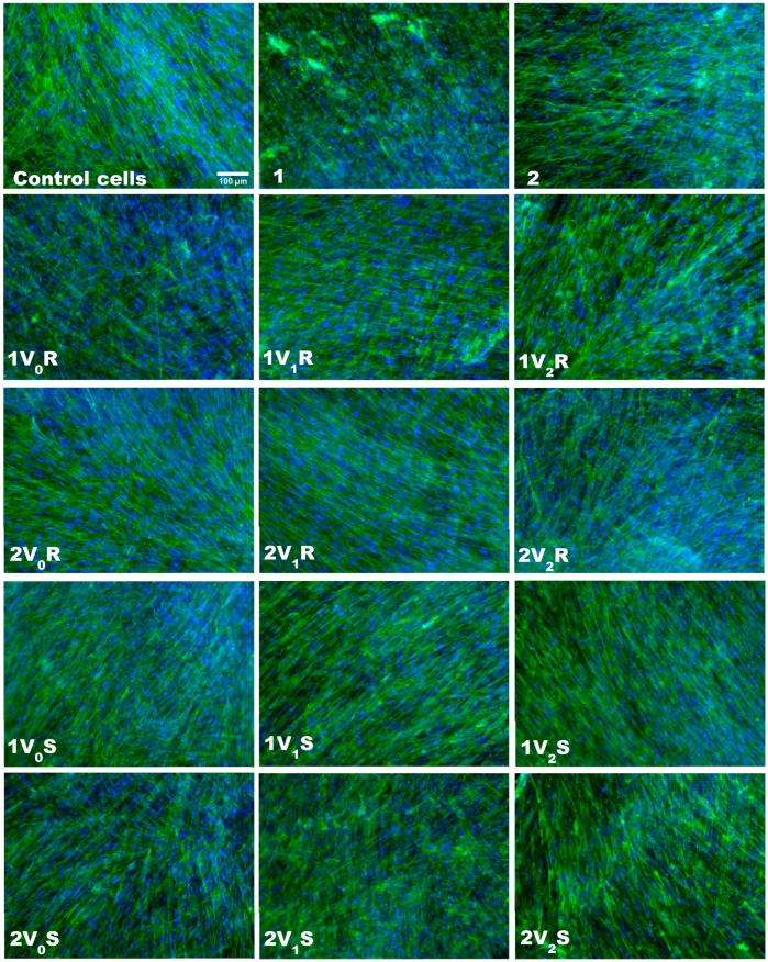
Figure 3.
Fluorescence images of actin filaments (green) and nuclei (blue) staining in human dermal fibroblasts cultured on inserts in the presence of fabrics treated in different experimental variants with sage (S) and rose (R) microcapsules for 12 h. Scale bar of 100 µm shown on first micrography is the same for all images.

