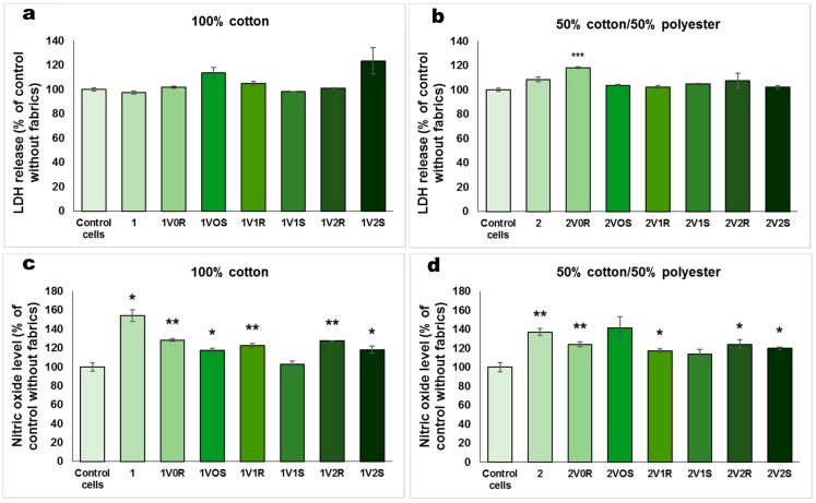
Figure 4.
Biocompatibility in terms of LDH release (a,b) and NO level (c,d) for 100% cotton and 50% cotton/50% polyester fabrics treated in different experimental variants with sage (S) and rose (R) microcapsules, that were incubated for 12 h with human dermal fibroblasts cultured on inserts. Data are represented relative to control cells without fabrics as mean ± standard deviation (SD) (n = 3). * p < 0.05, ** p < 0.01 and *** p < 0.001 compared to the control. In order to evaluate the biocompatibility of treated fabrics in a classic cell culture plate without inserts, the cell viability (Figure 5a,b), LDH release (Figure 5c,d) and NO level (Figure 5e,f) were assessed. In contrast with the good cell viability observed for the experiment performed in cell culture inserts (Figure 3), the results obtained after 12 h of exposure with textiles on the top of the cells showed a decrease in cell number compared to control. The cell viability measured for the materials functionalized by cationizing and padding was by 40–50% lower than for the control. Taking into account that the viability was even lower for the untreated 100% cotton fabric, it could be suggested that the functionalized textiles better protected the fibroblasts attachment on the surface of culture plate.

