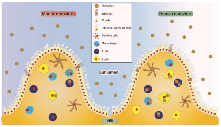
Figure 1.
Proposed tropism model of MNV and HuNoV to various cells in the human small intestine [36,50]. These viruses may enter in free form or as clusters [51] potentially crossing through microfold (M) cells [52,53] or through intestinal epithelial cells of the villus. Tuft cells express CD300lf and have been shown to be infected by MNV [39]. Norovirus can further cross the brush border of epithelial cells to infect cells of hematopoietic origin like dendritic cells [36,43], T cells [14,36,41], B cells [36,43], and macrophages [14,36,41]. All the cell types depicted as HuNoV positive have been shown to be positive for viral antigen or genome in either animal studies or human studies.

