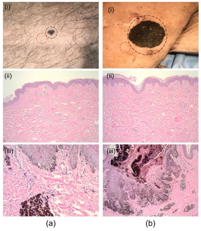
Figure 3.
Imaged suspect lesions. (a) (i) Abdominal, dark-brown pigmented plaque with irregular border confirmed as melanoma (black-circle), (ii) histology of nearby healthy skin (red-circle), and (iii) histology of the suspect lesion. (b) (i) Flank, large dark-brown plaque confirmed as melanoma (circled), (ii) histology of nearby healthy skin, and (iii) histology of the suspect lesion.

