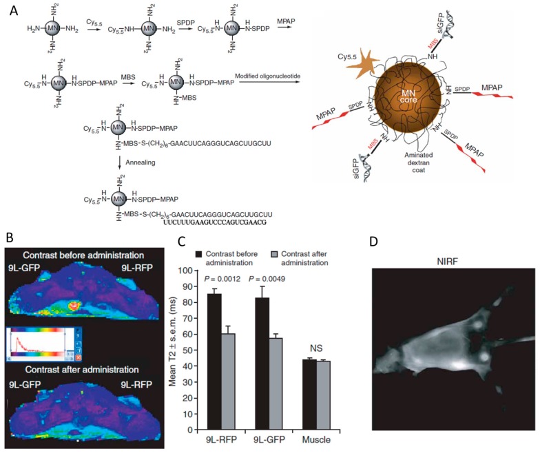
Figure 9.
(A) The synthesis of the magnetic nanoparticle (MN) by decorating Cy5.5, MPAP, peptide and siRNA step by step. (B) Magnetic resonance imaging of mice bearing bilateral 9L-GFP and 9L-RFP tumors before and 24 h after the administration of MN-siRNA conjugate. (C) The tumors exhibit a significant drop in T2 relaxivity, whereas the muscle tissue remained unchanged. (D) In vivo NIR imaging of the tumors in mice. Adapted with permission from [132].

