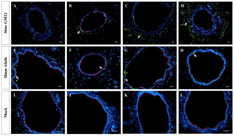
Figure 2.
Mycoplasma mycoides infection of caprine PCLS Caprine PCLS infected with Mmc GM12 (A–D), Mmm Afadé (E–H), and uninfected control (I–L). Slices were fixed after 24 h (A,E,I), 48 h (B,F,J), 72 h (C,G,K), and 96 h (D,H,L) p.i. Mmc GM12 colonizes the sub-bronchiolar tissue, and the invasion increased over time (A–D, white arrows). Mmm Afadé was mainly seen on the ciliated epithelial cells (E–H, white arrows). This indicates the difference in the tropism of both strains. There were no Mycoplasma cells in the uninfected control samples (I–L). Immunofluorescence images of tissue sections are shown, labeled with a polyclonal rabbit anti-Mmm PG1 antibody combined with a FITC-labeled goat anti-rabbit IgG secondary antibody (green) and a mouse monoclonal anti-β-tubulin-Cy3 antibody (red). Nuclei of caprine cells were counterstained with DAPI (blue). Scale bars: 50 µm.

