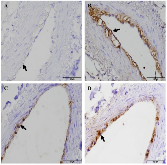
Figure 4.
Adherence of Mmc GM12 to caprine and bovine pulmonary endothelial cells. IHC showing endothelial cells stained with anti-Von Willebrand Factor antibody (a marker of endothelial cells) in caprine (A, black arrow) and bovine (C, black arrow) PCLS. Seriate sections were stained with anti-Mmm PG1 antibody to see adherent Mmc GM12 to the caprine (B, 96 hpi, black arrow) and bovine (D, 48 hpi, black arrow) endothelial cells in PCLS. Red- β-tubulin of ciliated cells, Green: Mmc, Blue: Nuclei of caprine cells. Scale bars = 50 µm.

