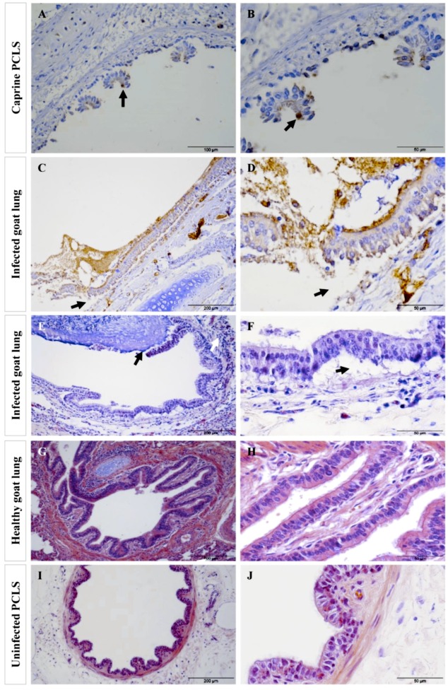
Figure 8.
Tissue destruction in caprine PCLS following infection with Mmc GM12 in PCLS and lungs of goats infected with Mmc GM12 in vivo. After 24 h of continuous infection, extensive detachment and destruction of the bronchiolar epithelial layer were observed in caprine PCLS infected with Mmc GM12 (A,B, IHC staining). Bacteria were mainly adherent to the ciliated cells (A,B, black arrows) and detach the ciliated cells 24 hpi leaving the basal cells. Similar histopathological changes were found in tissue sections of goat lungs experimentally infected with Mmc GM12 (C,D, IHC staining, E,F, H&E staining). Both the IHC overview (C, black arrows) and close up (D, black arrows) and H&E staining overview (E, black arrows) and close up (F, black arrows) show areas of detachment of the bronchiolar epithelial layer from the basement membrane region. In the in vivo samples, bacteria were highly adherent to the ciliated cells as observed by IHC staining of infected goat lungs (C,D). Infiltration of leucocytes was also observed in infected samples (E, white arrows). Histological section of an apparently healthy goat lung (G,H) and uninfected PCLS (I,J) revealed an intact bronchiolar tissue architecture. Scale bars: A = 100 µm, B, D, F, H, and J = 50 µm, C, E, G, and I = 200 µm.

