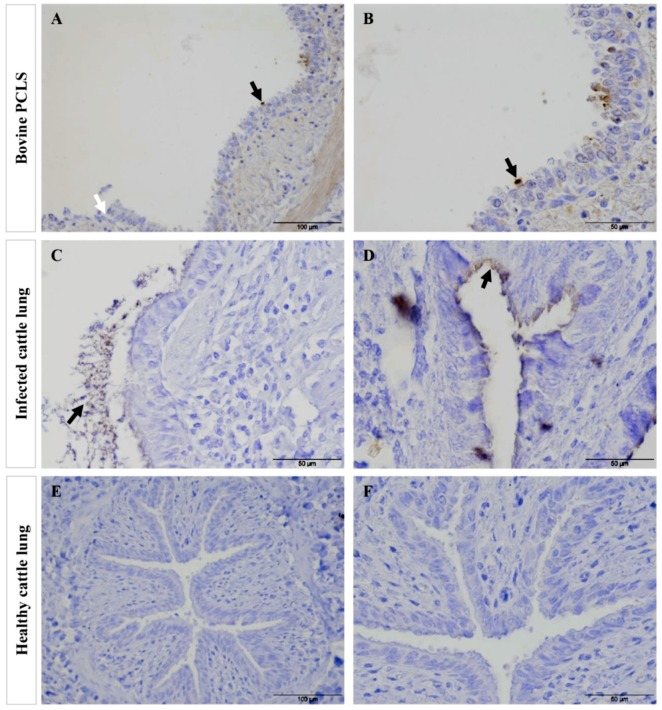
Figure 9.
Comparison of tissue destruction of bovine PCLS infected with Mmm Afadé versus lungs of cattle experimentally infected with Mmm Afadé (IHC). Bovine PCLS infected with Mmm Afadé (A,B), for continuous 24 h. Lungs of cattle infected with Mmm Afadé (C,D). Lung from healthy uninfected cattle (E,F). Bacteria were mainly adherent to the ciliated epithelial cells both ex vivo in PCLS (A,B, black arrows) and in vivo (C,D, black arrows). Destruction of the ciliated epithelial layer was observed in PCLS accompanied by detachment of the epithelial layer (A, white arrows) and partly in cattle infected with Mmm Afadé (C). Lungs from healthy cattle were also stained similarly and no mycoplasma was detected and revealed an intact bronchiolar tissue architecture (E,F). Scale bars: A and E = 100 µm, B, C, D, and F = 50 µm.

