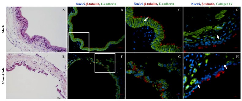
Figure 10.
Effects of 24 h infection with Mmm Afadé on the epithelial barrier in bovine PCLS. In uninfected control slices, the integrity of the epithelial barrier is demonstrated by H&E (A) and Immunofluorescence (B–D) staining of PCLS sections. Ciliated epithelial cells are connected by E-cadherin (B,C, white arrow) and the epithelial cell layer is attached on the Collagen IV containing basement membrane (D, white arrow). After 24 h of continuous infection with Mmm Afadé, the epithelial cell layer is detached from the sub-bronchiolar tissue (E), the connections between ciliated epithelial cells are partly broken (F,G) and the Collagen IV layer is largely degraded (H, white arrows). C and G are close-up views of the white rectangles on B and F, respectively. For IF images, PCLS sections were labeled with a monoclonal mouse anti-E-cadherin antibody or a polyclonal rabbit anti-collagen type IV antibody, combined with corresponding Alexa fluor 488 labeled secondary antibodies. Cilia (red) and nuclei (blue). Scale bars: H&E stains = 100 µm, B and F = 50 µm, D and H = 10 µm.

