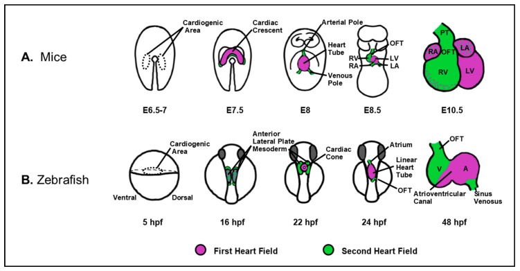
Figure 2.
Vertebrate cardiac development proceeds via the contribution of waves of differentiating progenitors, as depicted in mice (A) and zebrafish (B). During early heart development, cardiac progenitors are specified bilaterally within the lateral plate mesoderm and subsequently coalesce as the cardiac crescent in mice and cardiac cone, in zebrafish. In mice and zebrafish embryos, the FHF (magenta) first forms the cardiac tube followed by contribution of the SHF (green) to the poles of the developing heart. Subsequently, the heart undergoes an expansion process, including ballooning of the cardiac chambers, looping, and the generation of the septa in the case of a four-chamber heart. A: atrium; E: embryonic day; HPF: hours post-fertilization; LA: left atrium; LV: left ventricle; OFT: outflow tract; PT: pulmonary trunk; RA: right atrium; RV: right ventricle; V: ventricle.

