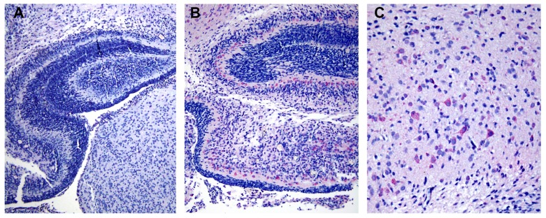
Figure 5.
Localization of envelope (E) protein in cerebral cortex of dengue virus (DV) serotype 2 infected mice. (A) Low magnification (10 ×) of mouse brain tissue from control group showed negative staining. (B) Positive E staining (pink) in cytoplasm of neuronal cells was revealed by anti-E specific monoclonal antibody (clone 4G2) and alkaline phosphatase secondary antibody at low magnification (10 ×). (C) A high magnification (20 ×) of B showed distribution of E protein in the majority of neuronal cells in the cerebral cortex region.

