Figure 1.
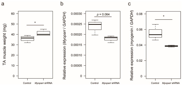
Knockdown of Myoparr attenuated skeletal muscle atrophy caused by denervation. (a) Box-and-whisker plots showing the weights of denervated tibialis anterior (TA) muscles of C57BL/6J mice electroporated with either control short hairpin RNA (shRNA) against LacZ or Myoparr-specific shRNA. TA muscle weights were measured 3 days post denervation (n = 4 per group). * p < 0.05, unpaired two-tailed Student’s t-test. Central black bar indicates median; lower and upper box limits are 25th and 75th percentiles, respectively; whiskers show maximum and minimum values. (b) The level of Myoparr expression in denervated TA muscles electroporated either with control or Myoparr shRNA (n = 3 per group) was measured by quantitative reverse transcription polymerase chain reaction (qRT–PCR). p = 0.064, unpaired two-tailed Student’s t-test. Data were normalized to glyceraldehyde-3-phosphate dehydrogenase (GAPDH) expression. (c) Quantitative reverse transcription–polymerase chain reaction (qRT–PCR) showing decreased myogenin expression by Myoparr knockdown 3 d post denervation (n = 3 per group). * p < 0.05, unpaired two-tailed Student’s t-test. Data were normalized to GAPDH expression.

