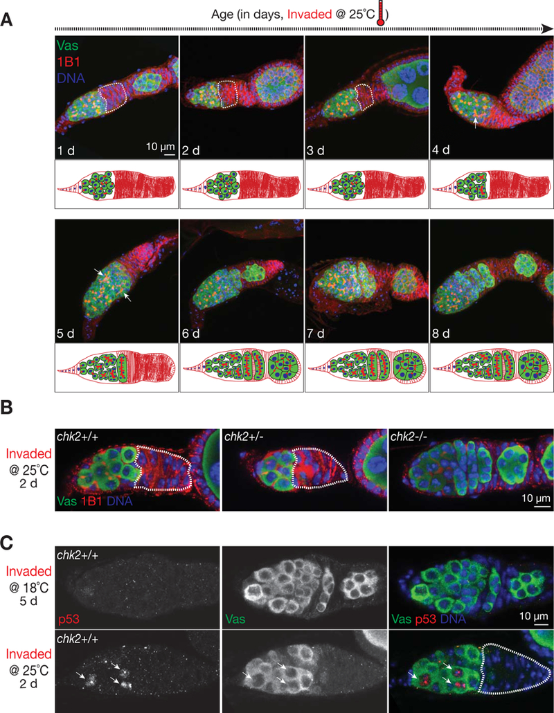
Figure 4. Germline stem cells silence the invading P-elements via the activation of Chk2/p53.

(A) Germarial structure from invaded progeny after vigorous P-element invasion. The germ cells are labeled by Vasa protein (green). The germaria are co-stained with 1B1 (red), an antibody that targets Hu-li tai shao protein that specifies germ cell stages in germarium and outlines cell membrane. Upon intensive invasion, the early stage germ cells are arrested, as evidenced by a “gap” region (circled by dash line) that contains no Vasa positive cells. Note that germ cells in the germarium display “dot” shaped structure from 1B1 staining, indicating that they are undifferentiated germ cells. Oogenesis reinitiates at day 4 or 5, as suggested by forming the “branch” structure from 1B1 staining (pointed by arrow).
(B) Germarial structure from invaded progeny that are either wild type (left), heterozygous (middle), or homozygous (right) for the chk2 gene. Mutating chk2 gene completely rescues the arrest phenotype, as evidenced by the germarium containing continuous germ cells and normal formation of cysts at different stages.
(C) p53 staining to detect the activation of DNA damage response. p53 signals (arrows) are readily detectable in the germarium from 25°C incubation.
