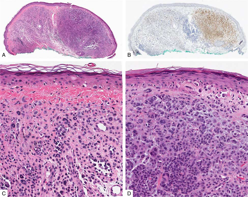
FIGURE 4.
Melanoma associated with a melanocytic nevus in the ear of a 63-year-old man. A, Nodular silhouette of the lesion with a more densely cellular tumor cell population on the right side of the lesion. B, IHC for PRAME stains only the densely cellular nodule. C, The less cellular area shows cytologic features of a melanocytic nevus. D, The PRAME-positive tumor cells are cytologically atypical. Cytogenetic analysis of the tumor cells revealed a number of chromosomal aberrations, including loss of 9p and gain of 8q (not shown).

