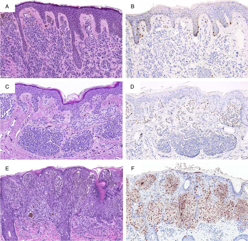
FIGURE 6.
PRAME immunoreactivity in nevi. A, Ordinary melanocytic nevus (H&E-stain). B, A few junctional melanocytes express PRAME (1+). C, Compound dysplastic nevus (H&E-stain). D, The center of the lesion contains a number of PRAME-positive melanocytes at the dermoepidermal junction and in the superficial dermis (2+). E, Predominantly junctional Spitz nevus on the cheek of a child (H&E-stain). F, The intraepidermal lesional melanocytes diffusely label for PRAME (4+).

