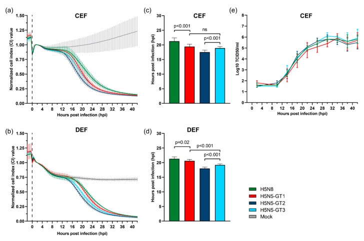
Figure 3.
Cytopathogenicity and replication of HPAI H5N5 viruses in primary avian cells. (a,b) Cytopathogenicity of highly pathogenic avian influenza (HPAI) H5N5 and H5N8 virus in primary chicken embryo fibroblast (CEF) and duck embryo fibroblast (DEF) cells measured by the real-time cell analysis (RTCA) system. The electrical impedance of the cell-covered electrodes was displayed as cell index (CI) value and normalized at two hours post infection (hpi). Virus was inoculated at a multiplicity of infection (MOI) of 0.001. Mock-infected cells were taken along as negative controls (grey). (c,d) The mean time at which the CI value decreased to 50% of the maximum (CI50) value after infection of primary CEF and DEF cells with HPAI H5N5 and H5N8 virus. The p-value was calculated using a two-tailed unpaired Student’s t-test with p < 0.05 considered statistically significant. (e) Growth curves of HPAI H5N5 and H5N8 virus in primary CEF cells. Virus was inoculated at a MOI of 0.001. Samples were taken at four hour intervals from 2 to 42 hpi and titrated to determine the medium tissue culture infective dose (TCID50) titres. Error bars indicate standard deviations (SD).

