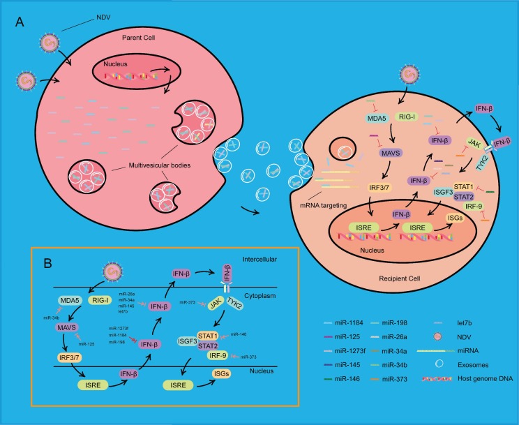
Figure 8.
Schematic showing that exosomes carrying miRNAs promote NDV infection. (A) During NDV infection, three miRNAs changed their expression and loaded into exosomes derived from multivesicular bodies (MVBs) of a NDV-infected HeLa cell, called the “parent cell”. MVBs fuse with the plasma membrane and release exosomes that contain these miRNAs. In another HeLa cell, called the “recipient cell”, these three miRNAs, transferred by exosomes, affect the IFN-β mRNA and further affect the expression of ISGs, eventually resulting in increasing NDV replication. (B) MicroRNAs may be involved in the IFN I pathway during NDV infection. MicroRNAs marked by the red arrow were found in the current research, and microRNAs marked by the pink arrow were found in other virus infections and remain to be elucidated.

