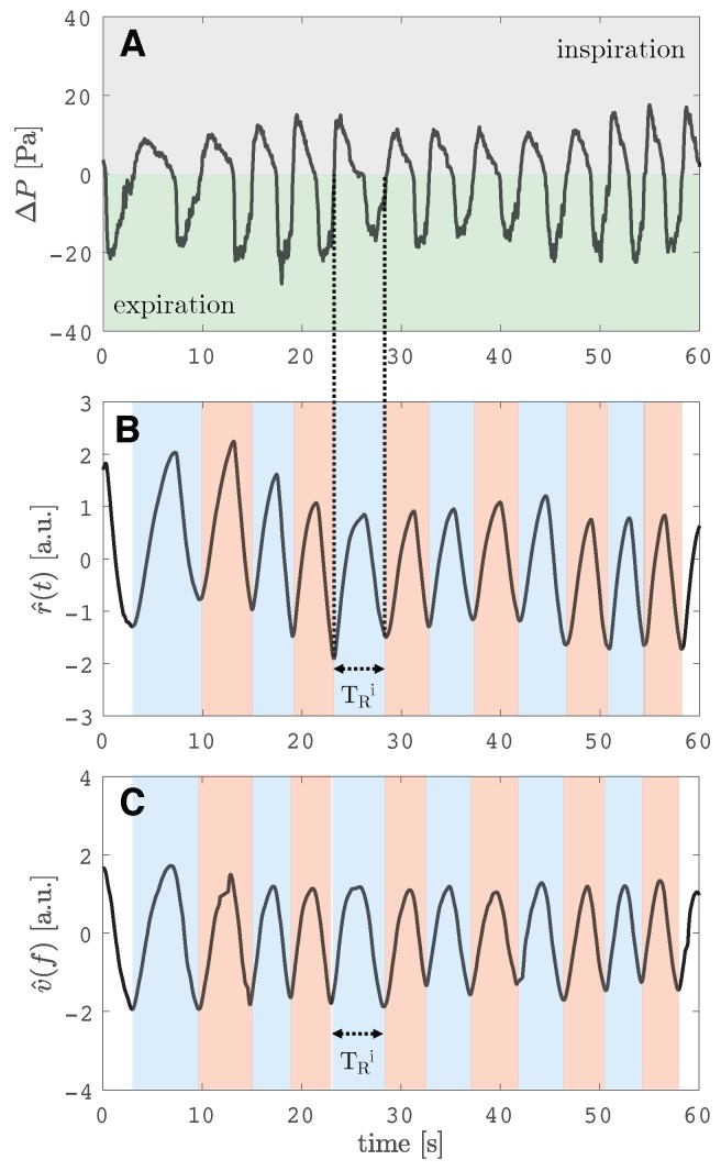
Figure 2.
(A) signal recorded by the reference instrument at the level of the nostrils. In grey, the signal collected during the inspiration (positive pressure), while in green, the signal recorded during the expiration (negative pressure). (B) Reference respiratory pattern signal () obtained from data processing of signal. (C) Respiratory pattern signal obtained from the proposed measuring system (). Figure in (B) and (C) show similar patterns: during the inspiratory phase the signal increases, while during the expiratory phase they decrease. The duration of one breath () is shown on both the and signals.

