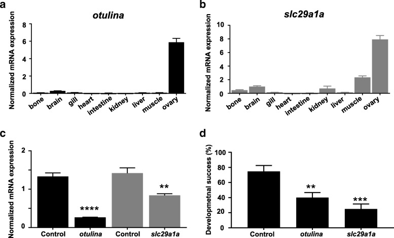
Fig. 3.
Tissue localization of otulina (a) and slc29a1a (b) based on qPCR assays. c: Expression level of otulina and slc29a1a in spawned eggs from mutant females mated with WT males as assessed by qPCR. 18S rRNA, beta-actin (bact), and elongation factor 1 alpha (EF1α) were used as internal controls, and experiments performed in triplicate. d: Developmental success in terms of survival rate of embryos at 24 h post-fertilization (hpf) from otulina- and slc29a1a-deficient mutant females mated with WT males. N = 4 r different females for otulina and N = 10 different females for slc29a1a, using eggs from at least three spawns for each individual female

