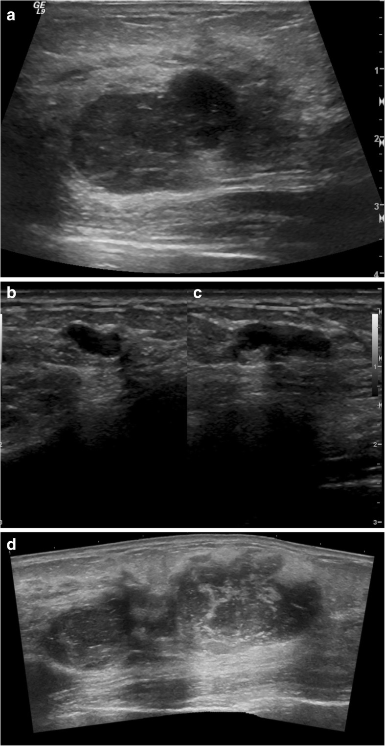
Fig. 1.

a Ultrasound image of left breast antiradial plane at 1:00 position 9 cm from the nipple demonstrates a mixed cystic and solid, irregularly shaped, hypoechoic, and heterogeneous mass with ill-defined margins and areas of posterior acoustic enhancement. Overall lesion size is 3.7 × 2.2 × 4.0 cm. b Ultrasound image of left breast antiradial plane at 12:00 position 8 cm from the nipple demonstrates a predominately solid, lobulated, hypoechoic, and heterogeneous mass with smooth margins and only minimal posterior acoustic enhancement. Overall lesion size is 3.4 × 1.8 × 2.7 cm. c Ultrasound image of left breast split screen antiradial (left) and radial (right) planes in 2:00 position 6 cm from the nipple demonstrates a predominately solid, lobulated, homogeneous, and hypoechoic mass with smooth, well-defined margins and areas of posterior acoustic enhancement. Overall lesion size is 0.8 × 0.4 × 1.2 cm. d Ultrasound image of the left breast with panoramic view shows the lesions shown in a and b may coalesce into a single larger lesion. This coalescent lesion is congruent with the pathologic findings of an 8.8-cm lesion
