Fig. 1.
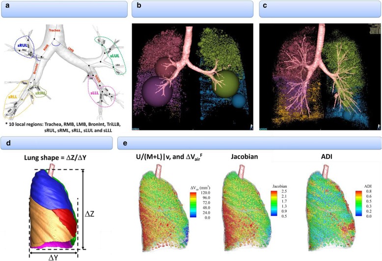
An expanded set of imaging-based metrics including emphysema percentage, tissue fraction at TLC and RV. a Inspirational image-based local structures: θ, Cr, WT*, and Dh*. b Expiration image-based global and lobar function: AirT%. c Inspiration image-based global and lobar function: Emph%. d Global structure:. e Registration-based global and lobar functions:.

