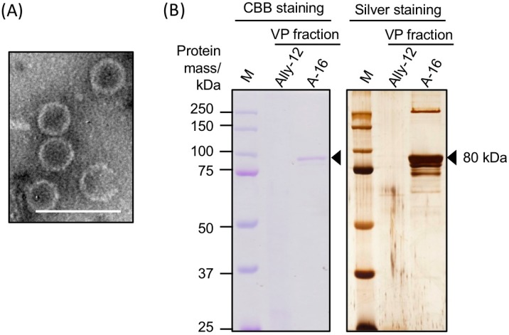
Figure 3.
Components of purified virus particles of AalVV1. (A) Transmission electron micrograph of negatively stained particles of AalVV1. The white scale bar denotes 100 nm. (B) Structural proteins included in the VP fraction of A. alternata strain Ally-12 and A-16. Proteins were denatured in modified Laemmli’s sample buffer containing 0.6% 2-mercaptoethanol and electrophoresed in 10% polyacrylamide gels. The gels were stained with Coomassie Brilliant Blue R-250 (CBB) or silver nitrate. The lane M shows approximate protein molecular size with the Precision Plus Protein Dual Color Standards (Bio-Rad Laboratories, Inc., Hercules, California, USA). The black arrows are pointed to the major 80 kDa protein band subjected to LC-MS/MS analysis.

