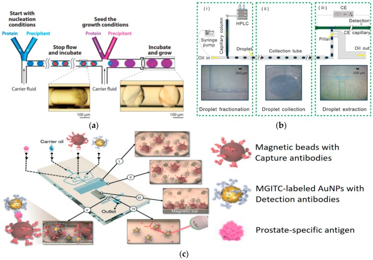
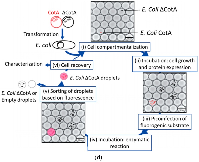
Figure 4.
Droplet-based microfluidics for proteomics. (a) Separating the stages of nucleation and growth in the crystallization of “severe acute respiratory syndrome (SARS) protein” yields single crystals, reproduced with permission from [62], published by John Wiley and Sons, 2006. (b) A droplet-interfaced 2D nano liquid chromatography-capillary electrophoresis (LC-CE) system. The HPLC effluent is fractionated into a series of nanoliter droplet units right after chromatography (panel (i)), and collected and stored in a tube (panel (ii), tube inside diameter: 0.38 mm), before drop-wise analyzed in capillary electrophoresis (CE) (panel iii), reproduced with permission from [72], published by Elsevier, 2015. (c) Schematic illustration of the surface enhanced raman scattering (SERS)-based microdroplet sensor for wash-free magnetic immunoassay, reproduced with permission from [77], published by Royal Society of Chemistry, 2016. (d) A flexible droplet-based micro-fluidic platform that can be used for high-throughput screening of Escherichia coli cells for measuring the CotA enzymatic activity, reproduced with permission from [84], published by Royal Society of Chemistry, 2014.


