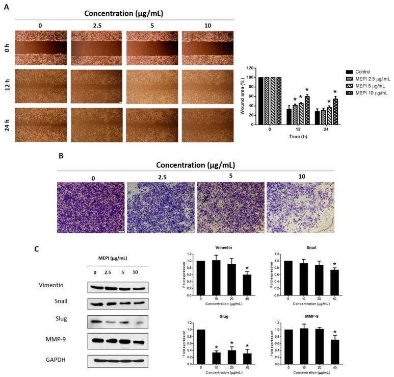
Figure 2.
MEPI inhibited the epithelial–mesenchymal transition (EMT) in MDA-MB-231 cells. (A) Migration of MDA-MB-231 cells as determined by wound-healing assay. Wound area was calculated from a representative of at least three independent experiments. Percentages of wound closure at the indicated time points after MEPI treatment are shown. (B) Invasion of MDA-MB-231 cells. After incubation with MEPI for 24 h, cells that migrated through the Matrigel were stained with crystal violet and visualized by phase-contrast microscopy (40×). (C) Western blot for Vimentin, Snail, Slug, and MMP-9 after treatment for 24 h with MEPI at the indicated concentrations using GAPDH as the internal control. Band intensities were measured using ImageJ software. Data are means ± standard deviation (SD). * p < 0.05.

