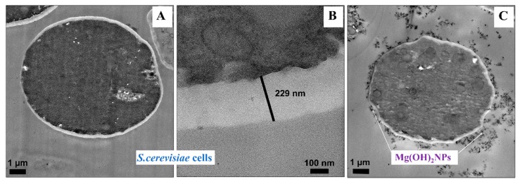
Figure 5.
Transmission electron microscopy (TEM) images of S. cerevisiae after being incubated for one day with bare Mg(OH)2NPs: (A) A control sample without Mg(OH)2NPs. (B) A high-resolution TEM image of the S. cerevisiae wall without Mg(OH)2NPs. (C) S. cerevisiae cells incubated with 1000 µg mL−1 Mg(OH)2NPs showing the attachment of Mg(OH)2NPs to the outer cell surface.

