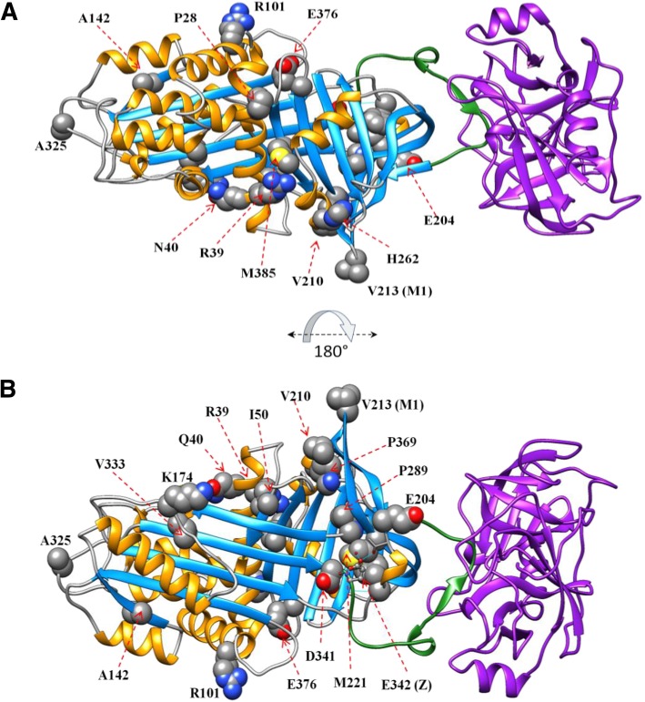
Fig. 1.
Structure of AAT indicating the location of missense residues. The AAT protein (PDB code 1OPH) is shown in ribbon representation coloring according to secondary structural elements (alpha helices shown in orange, beta strands shown in light blue), and the position of missense changes showing the wildtype residue in sphere representation and labeled with the residue name and position. The purple ribbon protein is trypsinogen. The stretch of amino acids that comprise the reactive center loop are shown in green ribbon representation. A = front view; B = rear view (rotated 180 degrees about the x-axis). AAT, Alpha 1 Antitrypsin

