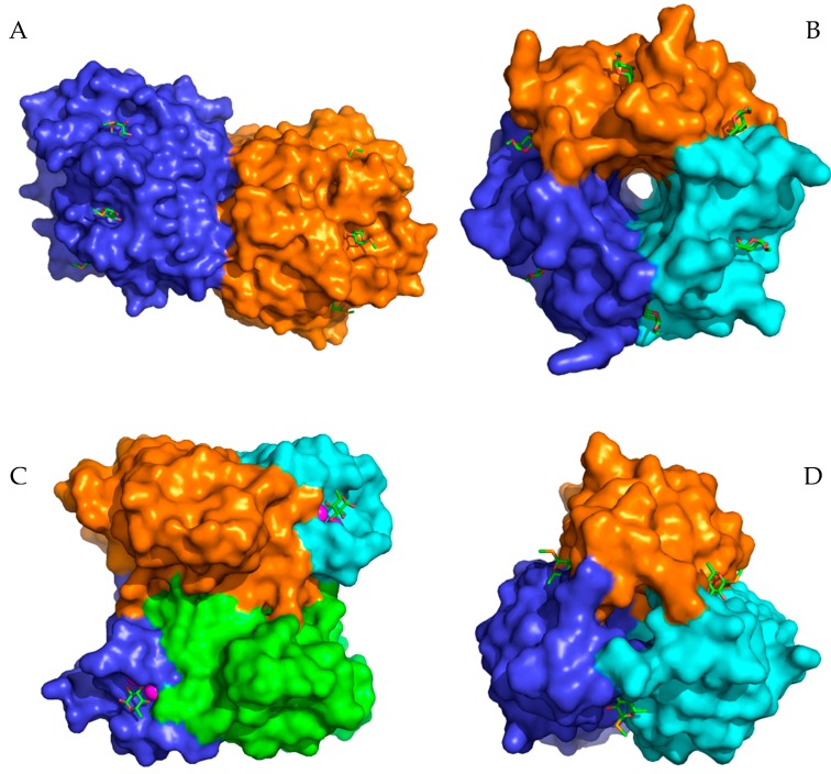
Figure 1.
Structures of selected lectins from pathogenic microorganisms used for the inhibition studies. (A) AFL dimer in a complex with seleno fucopyranoside (PDB 4AGI). (B) RSL trimer in a complex with methyl α-l-fucopyranoside (PDB 2BT9). (C) PA-IIL tetramer in a complex with α-l-fucopyranoside (PDB 1UZV). (D) Trimer of the BC2L-C N-terminal domain in a complex with seleno fucopyranoside (PDB 2WQ4). Carbohydrates in the binding sites are depicted as sticks. The magenta spheres represent calcium ions in the binding sites of lectin PA-IIL.

