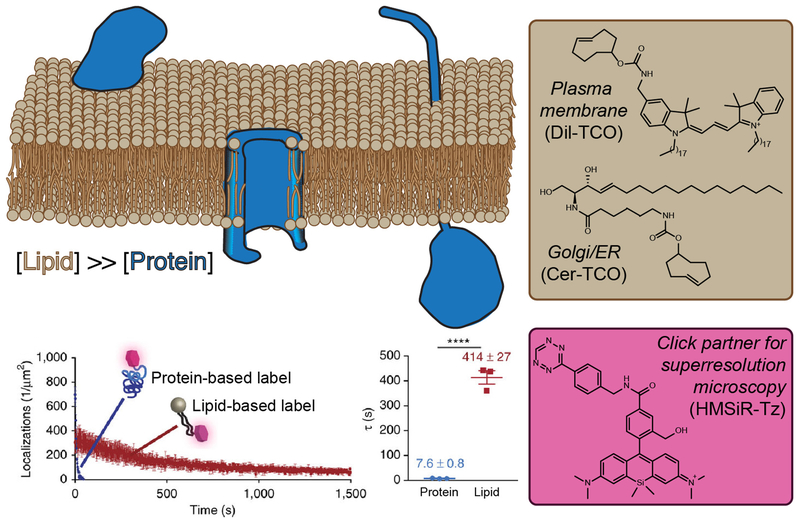
Figure 3. Targeting Lipids Enables Time-Lapse Super-Resolution Microscopy Visualization of Organelles.
(A) Lipids (tan) have a higher molar density than proteins (blue) within a given surface area of a membrane bilayer and, therefore, intracellular membranes can be densely tagged with lipid probes bearing reactive handles capable of rapid ligation with a flA) Lipids bearing the opposing click partner. (B) Clickable lipid-based probes for targeting specific organelles. (C) An appropriately functionalized click partner bearing a hydroxymethylene silicon rhodamine dye. This probe switches between nonfluorescent and fluorescent forms within a hydrophobic environment, enabling the extended imaging times relative to identically labeled proteins. Graphs show photobleaching time course (D) and fluorescence half-life (E) of lipid and protein probes targeted to the same organelle and bearing the same silicon rhodamine dye.

