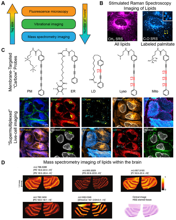
Figure 4. Less Is More: Non-Fluorescence Methods for ‘Supermultiplexed’ Imaging with Smaller Tags.
(A) Comparison of fluorescence imaging with vibrational imaging and mass spectrometry imaging in terms of tag size and number of species that can be detected simultaneously (‘colors’). (B) Stimulated Raman spectroscopy (SRS) imaging can be used to visualize lipids by exciting the vibrational modes of the CH2 groups within lipid tails (left) without any exogenous labels. Deuterated palmitate allows for pulse-chase imaging of the subset of lipids containing this particular lipid tail (right). (C) SRS imaging can be combined with fluorescence microscopy to enable ten-color imaging in live cells. Multiple vibrational ‘colors’ are created by extending the length of the dyes in polyyne chain and by introducing 13C–12C pairs into the chain, indicated in red. Such probes are dubbed ‘carbow’ (for carbon rainbow) and can target different organelles, including the plasma membrane (PM), endoplasmic reticulum (ER), lipid droplets (LD), lysosomes (lyso), and mitochondria (mito). (D) Mass spectrometry enables label-free imaging of lipids within tissues. Shown are images of phosphatidylethanolamine (PE), phosphatidylglycerol (PG), phosphatidylinositol (PI), phosphatidylserine (PS), and sulfatide (SHexCer) within a brain section taken from a mouse.

