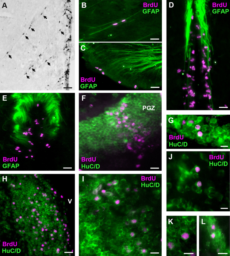
Figure 6.
Migration and differentiation of BrdU-labeled cells in the brain of male A. burtoni. A: Elongated BrdU-labeled nuclei (arrows) migrating away from the midline ventricular proliferation zone (on right) in the periventricular nucleus of the posterior tuberculum (TPp) at 1 day post injection. B: GFAP-labeled radial glial fiber guiding BrdU cells in the TPp. C: GFAP radial glial fibers extending to the proliferation zone in the preoptic recess of the PPa. D: GFAP-labeled glial fibers along the midline near the diencephalic proliferation zone in the region of TPp. E: GFAP fibers near BrdU-labeled cells in the NRP of the hypothalamus. F: BrdU-labeled cells in the periventricular gray zone (PGZ) of the dorsal tectum are colabeled with the neuronal marker HuC/D. G: Proliferating cells that express HuC/D in the ventromedial thalamic nucleus. H: Double-label of BrdU-labeled cells and HuC/D in the ventral nucleus of the ventral telencephalon (Vv) along the midline ventricle (V). I: Example of HuC/D-expressing BrdU-labeled cells in the preoptic area. J–K: Higher magnifications of BrdU-labeled cells colabeled with HuC/D in the lateral part of the dorsal telencephalon (Dl) (J), preoptic area (K), and vagal lobe of the rhombencephalon (L). Representative photomicrographs in B–L are of coronal sections at 30 days post BrdU injection to show expression of BrdU-labeled cells (magenta) with the radial glial marker GFAP (green; B–E) or the neuronal marker HuC/D (green; F–L). For abbreviations, see list. Scale bar = 50 μm in A,C–F,H,I; 20 μm in B,G,J–L.

