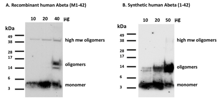
Figure 7.
Western blot characterization of the recombinant human Aβ(M1-42) peptide. A. 10, 20, and 40 μg of the recombinant Aβ(M1-42) peptide was run on an SDS-PAGE gel and bands visualized using the 6E10 antibody with Western Blot. Lower concentrations of 10 and 20 μg show monomeric bands at 4 kDa while the 40 μg lane shows oligomeric bands at 14–17 kDa along with the monomeric band. All 3 concentrations show a slight amount of high molecular weight bands at 38–49 kDa suggesting a few aggregated forms of the peptide in the mixture. B. Synthetic human Aβ1-42 for reference shows similar monomeric bands at 10, 20, and 50 μg concentrations. More oligomers are present in the synthetic peptide.

